M
Leave a Legacy of Giving
You can support our mission of changing lives by saving sight in multiple ways!
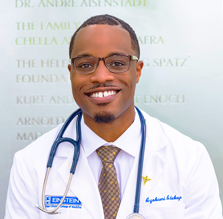
In recent years, advancements in medical research have pushed the boundaries of what was once considered feasible in the field of ophthalmology. One such study observed the effect of the expansion of eye bank donor age on corneal endothelial cell density and surgeon acceptance rate of those tissues. Donor characteristics, endothelial cell density, and acceptance of tissues for use in surgery were compared between age groups in five-year intervals. The aim was clear: to assess how this expansion would influence the availability of corneal tissue for transplantation and challenge existing biases against older donors. Saving Sight was proud to partner with Dr. Roy Chuck and Ayobami Adebayo, among other researchers, to provide the corneal tissue used during this study.
This single-site study featured 25,969 corneas from eye bank donors from 2018 – 2022 between the ages of 2 and 75. At the beginning of 2022, the donor age limit was increased to 80 years old, thus allowing donated tissue from older donors to be used in transplants. The age limit increase allowed 411 more cornea donations, which led to 208 more transplants. The average endothelial cell density for the 71-75 age group was 2,349 cells/mm2, compared to 2227 cells/mm2 in donors aged 76-80. The difference of 122 cells/mm2 doesn’t seem like much, but the study saw that donors aged 71-75 had a 38% surgeon rejection rate, while those aged 76-80 had a 48% surgeon rejection rate.
There could be multiple reasons for the difference in surgeon rejection rate, but one could be age bias. Traditionally, corneas that come from older donors are looked at as not viable, but this study showed that corneas from older donors are still very viable for transplant. While there isn’t a shortage of corneas for transplant in the United States, there is a global shortage, resulting in patients in other countries waiting on the transplant list for months.
“If we can expand the age pool to increase and even get some more donations, that will increase transplants,” Adebayo said. “And I think there’s just a lot more room for growth in that area.”
This study is important for the future of ophthalmology and eye banks because there is a cornea shortage in other countries. By increasing the donor age limit, more corneas will be available for transplant globally. Expanding the donor age limit and using those tissues in surgery can give the gift of sight to many more people in need across the globe. Potential age bias isn’t the only factor in the difference in surgeon rejection rate, but it does show that more work needs to be done.
“So, no matter what you do, there’s still a bias against age and many things that we do in life, and that bias never disappears,” Chuck said. “…We can do one of two things. We can work to death to change everyone’s mind just by talking to them. It’s very difficult to do, and it’s much easier for us to generate data, and that’s what we’re doing. That’s the whole basis of research, you know, change thought by proving it with data.”
As researchers navigate the complexities of eye, organ, and tissue donation and transplantation, they remain committed to ensuring equitable access to sight-restoring treatments for all. Increasing the donor age limit will increase viable donor tissue for transplant, allowing those in underserved areas to receive the gift of sight. The journey to redefine how corneal donations are handled isn’t solely about science; it’s a moral duty with the chance to change lives by saving sight.
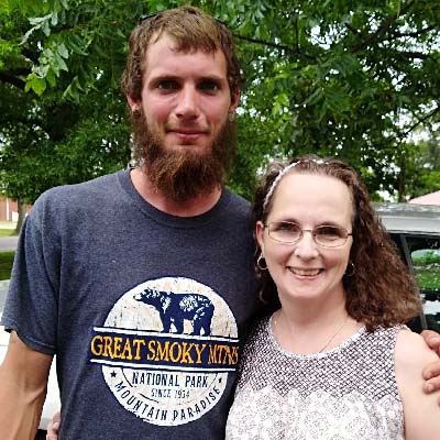
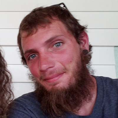
“To know Tommy, he preferred Thomas, was to know someone who was helpful, respectful, loyal, and enjoyed spending time with his family, friends, and his dog. Thomas was happiest working with his hands and going hiking, camping, and riding his motorcycle,” says his mother Charlotte.
Thomas had made the decision to join the organ, eye and tissue donor registry. “I didn’t know until after the accident that he was an organ donor, but I wasn’t surprised,” says Charlotte. “Through his cornea donations, his legacy in helping others continues so that he could keep on giving to others. He didn’t know a stranger; he was that kind. Because Tommy’s corneas were separately donated, he was able to be doubly generous.”
Thomas & his mother Charlotte
Thomas & his brother Colt in Colorado
Thomas 2018
His left cornea helped restore the eyesight of a cornea transplant recipient and his right cornea went to a research facility in order to help discover the cause, and hopefully a cure, for a genetic eye disease called PPCD (Polymorphous Corneal Dystrophy).
Charlotte took part in Saving Sight’s correspondence program to say thank you to his recipients and to share about Tommy.
“I reached out to Tommy’s left cornea recipient to build a connection. I wanted to know who had been blessed with the gift of sight because of my oldest son’s generosity. The recipient happily replied. And we began to learn – I learned about the recipient as a person and the recipient was learning about Thomas as a person. It has been very rewarding for me.”
She also chose to reach out to the researcher who received Thomas’ right cornea tissue. “Because one of Tommy’s corneas went to a research facility, I wanted to make sure the scientists were making the most of their precious and life-giving donation. Eye research is important to me because it impacts the future. If PPCD (Polymorphous Corneal Dystrophy) can come even one step closer to being cured, then it’s worth the effort. If one person doesn’t have to worry about possibly going blind, then eye, tissue, and organ donation are worth giving.”
As a donor family, Charlotte has found comfort in supporting donation and in Thomas’ legacy living on. “As a donor family, donation is a way to give back, the ultimate in paying it forward. Finding out that my oldest son wanted to give of his self after he was gone, made me even more proud to be Thomas’ mom. Because I honored my son’s wish, he lives on – twice. I am comforted that Thomas’ donation benefited a recipient to see clearly again and research is actively exploring a cure for PPCD.”
“Thomas made a conscious decision to give of his self beyond his last breath. And I could not be more proud of him for his ever continuing gift of sight. Thomas, my oldest son, had the most deep blue eyes that continue to see even though Thomas cannot.”
Joining the Organ Donor Registry
You can join the national organ, eye and tissue donor registry at registerme.org. “I have been considering becoming an organ donor. Even though I wear glasses, I have been advised that I could still give the gift of sight to someone else,” says Charlotte.
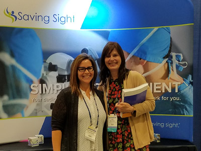
The American Academy of Ophthalmology (AAO) annual meeting was held in San Francisco this October. This meeting is the world’s largest association of eye physicians and surgeons, bringing together leaders from around the world.
Our Saving Sight Team attends each year to learn about new trends in eye banking and ophthalmology, and to let our partners know innovations we are exploring. Attending also allows us to be a resource to our partners in sharing information to those who couldn’t attend or might have missed part of the meeting. This experience allows us to learn together side-by-side and be a stronger support to our partner surgeons in their work.
In case you missed AAO, or if you want a refresh, here are our top takeaways from the conference and presentations:
Kansas City, Mo., August 21, 2019 – Jody Summers, PhD, professor of cell biology at the University of Oklahoma Health Sciences Center, studies the extracellular matrix of the sclera and changes associated with myopia development. Myopia is largely regulated by the visual environment. Through her studies, she hopes to understand why myopia interacts the way it does so intervention is possible.
“Nearly 50 percent of the world’s population will be myopic by 2050,” says Dr. Summers. “In some countries, such as those in East Asia, myopia is the leading cause of blindness. It can be completely preventable with how the eye responds to the visual environment if we can learn why.”
As a postdoctoral fellow in ophthalmology, Dr. Summers identified that there was little research with regard to the sclera. “Through research, I discovered that the sclera is not just a static container in the eye. The sclera is actually very active and can alter its compositions to control the refractive state or length of the eye. What happens in myopia is the sclera begins to elongate. Because sclera is a very responsive tissue, we are trying to understand how sclera remodeling happens so we can work to regulate it – which has taken us to this study.”
Saving Sight was honored to be a co-author on a research project at the 2017 Annual Meeting of the Association for Research in Vision and Ophthalmology (ARVO) with Dr. Summers, titled Isolation and Transcriptome Analyses of Choroidal Retinaldehyde Dehydrogenase-2 (RALDH2) Expressing Cells.
At ARVO 2017, Dr. Summers successfully isolated cells in the choroid (the vascular layer of the eye, located between the sclera and the retina) of both chick and human eyes which have been found to produce the enzyme retinaldehyde dehydrogenase 2 (RALDH2).
“The ARVO abstract was preliminary research to see if we could isolate those cells,” says Dr. Summers. “We first had a pilot study where Saving Sight provided us with two, reduced fee whole globes. This showed we could isolate the retinoic acid in the choroid.”
In summer 2019, Dr. Summers received National Eye Institute at NIH funding to further study the isolated cells through transcriptome analyses. Part of that grant contributes to the fees associated with receiving 10 whole globes or poles (which is the whole eye minus the cornea) from Saving Sight. “This next step is to do it again and do the transcriptome analyses to identify the cells in the choroid that make the retinoic acid.”
Dr. Summers adds that retinoic acid is a powerful chemical and they are interested in identifying the cells that produce it. “If we identify these cell types we can, perhaps, use that information to develop strategy to control the synthesis of retinoic acid,” she says.
Since the human genome has been sequenced, Dr. Summers will be able to sequence the cells from the choroid and compare with the human genome sequence to gather more information as to which cell type is responsible for retinoic acid synthesis.
Collaboration between Researchers and Eye Banks
“I value working with Saving Sight because it was very easy to get the collaboration set up,” says Dr. Summers. “I love that they are interested in research and feel like it’s a two-way relationship. I’m really glad they are available and willing to introduce a new technique or procedure that is compatible for research.”
Dr. Summers adds that one of most valuable connections in eye banks and researchers working together is the ability for Saving Sight to provide human eye tissue for research. “NIH funded research hopes to help humans through the betterment of health. Being able to provide tissue like Saving Sight does under the conditions needed is of value because not many eye banks do that.”
About Saving Sight
Saving Sight is a nonprofit eye bank with a mission to change lives by saving sight. Founded in 1960, Saving Sight has grown to become one of the nation’s leading eye banks and is focused on providing innovative solutions to its clinical and research partners. Saving Sight welcomes customized research collaborations that meet your research tissue needs and strives to advance the field of corneal transplantation through dynamic collaborations with medical centers and researchers. Headquartered in Kansas City, Mo., Saving Sight facilitates eye donation in Missouri, Kansas, and Illinois, serving transplantation and research specialists locally and around the world.
Click here to learn more about Saving Sight’s customized research collaborations.
Previous studies in the field of corneal transplantation have determined the importance of endothelial cell density in the health of corneal tissue grafts in terms of graft success rate. According to new research presented this week at the 2019 Annual Meeting of the Association for Research in Vision and Ophthalmology (ARVO) in Vancouver, differences in the storage media solutions can have an effect on endothelial cell density.
Through the study, a retrospective analysis was conducted on a donor database from the Kansas City-based nonprofit Saving Sight to compare eye bank donor corneal endothelial cell density (ECD) after storage in Optisol GS and Life4°C solutions. The data analyzed included 24,581 donated eye bank corneas from 2011 through 2017 stored in Optisol GS or Life4°C solutions.
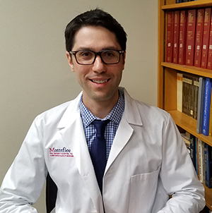
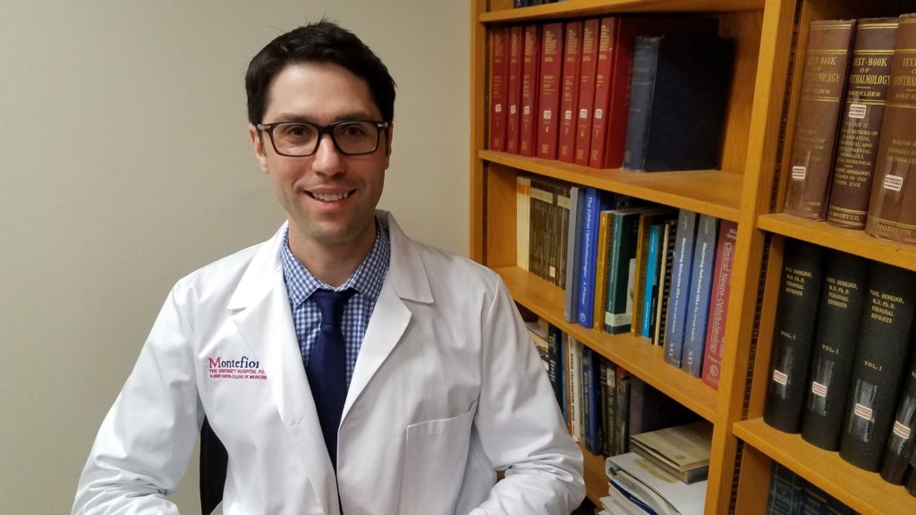
This project was led by Daniel Polla, MD, Ophthalmology Resident at Montefiore Medical Center and the Albert Einstein College of Medicine Department of Ophthalmology and Visual Sciences, in collaboration with Saving Sight.
“My project looks at the different storage media solutions that donor corneas were stored in from 2011 to 2017 at Saving Sight. The preliminary findings demonstrate a small difference in endothelial cell density after storage in Optisol GS or Life4°C, with a higher ECD after storage in Optisol GS. The differences found between donor corneas in each of the storage media groups may be due to factors that were not accounted for over time such as changes in processing protocols, equipment used to measure ECD, and variability in specular microscopy. While it is possible that Optisol GS better preserves the endothelium, one potential cause for the small difference in ECD between groups is variation in endothelial cell visualization during specular microscopy due to differences in solution color,” said Dr. Polla.
“This research is important to the field of ophthalmology and corneal transplantation because it may influence the way that corneas are stored and/or evaluated prior to transplantation, ultimately leading to better graft success rates and outcomes for patients.” Dr. Polla added.
Saving Sight Chief Business Development Officer Patrick Gore, RN, CEBT, Director of Business Development Lynn Forest-Smith, and Chief Operating Officer Tina Livesay were co-authors on the study. As an eye bank that facilitates eye donation for transplant and research, the Saving Sight team is proud to support this project and to work in collaboration with Montefiore and Drs. Polla, Rand and Chuck.
“We collect a large amount of data on all of our donor cases and corneas as a part of normal operations. This was a great opportunity to have these data parameters analyzed collectively for this study. As an eye bank, this allows us to help researchers advance the field of corneal transplantation to help honor the gift of sight through better recipient outcomes,” said Tina Livesay, Saving Sight Chief Operating Officer.
This study highlights the importance of collaboration between eye banks, medical centers and researchers in advancing the fields of corneal transplantation and ophthalmology.
Abstract:
An analysis of donor corneas stored in Optisol GS and Life4°C solutions
Authors: Daniel J. Polla MD, Gabriel M. Rand MD, Patrick K. Gore RN CEBT, Lynn Forest-Smith CEBT, Tina Livesay CEBT, Roy S. Chuck MD PhD
Dan Polla, MD, Ophthalmology resident at Montefiore Medical Center, details his latest research study with Saving Sight that he will present at ARVO 2019.
According to new research presented this week at the 2019 Annual Meeting of the Association for Research in Vision and Ophthalmology (ARVO) in Vancouver, several potential risk factors, including donor diabetes mellitus, can lead to damage during Descemet Membrane Endothelial Keratoplasty processing.
There are limited studies identifying risk factors for damage when processing Descemet Membrane Endothelial Keratoplasty (DMEK), which has a high processing failure rate as compared to Descemet Stripping Automated Endothelial Keratoplasty (DSAEK). Through a study with lead researcher Gabriel Rand, MD, second year ophthalmology resident at Montefiore Medical Center and the Albert Einstein College of Medicine Department of Ophthalmology and Visual Sciences and Saving Sight, an analysis of potential risk factors was performed. Collaborative research partner, Saving Sight performs a high volume of DMEK processing.
Through the study, a retrospective analysis was completed with logistic regressions on all 385 DMEK tissues processed from 319 eligible donors at Saving Sight from July 2014 to June 2017. The study concluded that eye bank DMEK processing has significant rates of damage and risk factors for processing failure including donor diabetes mellitus, individual technician ability and the technician learning curve.
“Myself, Dr. Chuck and a bunch of us here at Montefiore have been working with Saving Sight to prepare an abstract for ARVO this year,” said Dr. Rand. “The topic of the abstract is the effect that donor diabetes status has on the corneal transplant quality. This is a really important subject because diabetes is a disease that is increasing in prevalence every year and it’s a multisystem disease – it affects every single part of the body, the cornea notwithstanding. There’s really not a tremendous amount of literature on what the potential influences that donor diabetes status has on these cornea transplant tissues. So what our contribution is, is to add to the fund of knowledge and to really examine does a donor diabetes status affect endothelial cell health, does it affect preparation failure rates, things like this, so it’s been a really exciting research project for us.”
Saving Sight Chief Business Development Officer Patrick Gore, RN, CEBT, Director of Business Development Lynn Forest-Smith, and Chief Operating Officer Tina Livesay were co-authors on the study. As an eye bank that facilitates eye donation for transplant and research, the Saving Sight team is proud to support this project and to work in collaboration with Montefiore and Drs. Rand and Chuck.
“It is a privilege for me to work with such a devoted team at Montefiore who want to advance the science of ophthalmology,” said Lynn Forest-Smith, Director of Business Development at Saving Sight. “I have a real passion for the work we do at Saving Sight and teaming up with these folks allows us to take our findings back to the eye banks, apply what we learn to improve our services and ultimately, help more people see.”
This collaborative study helps advance the fields of corneal transplantation and ophthalmology between eye banks, medical centers and researchers and helps identify factors that contribute to better grafts and better outcomes for patients.
Abstract:
Factors associated with eye bank descemet membrane endothelial keratoplasty processing damage
Authors: Gabriel M. Rand MD, Patrick K. Gore RN CEBT, Lynn Forest-Smith CEBT, Tina Livesay CEBT, Roy S. Chuck MD PhD
Gabriel Rand, MD, second-year Ophthalmology resident at Montefiore Medical Center, gives an overview of his recent research collaboration with Saving Sight for ARVO 2019